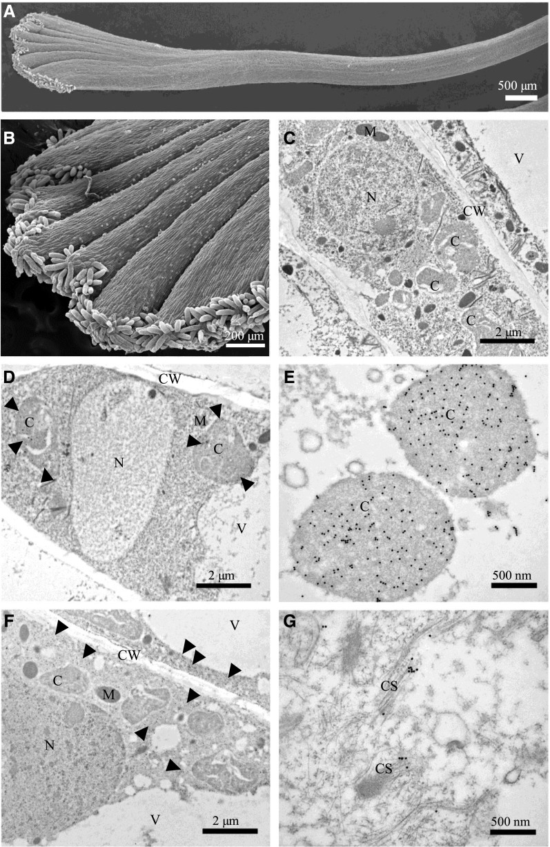
Figure 7.
Subcellular localization of CsCCD2 and CsUGT74AD1 in C. sativus stigmas. A and B, Scanning electron microscopy images of C. sativus stigmas. C to G, Immunoelectron microscopy (IEM) of C. sativus stigmas. IEM reactions were performed with an anti-CsCCD2 antibody (D and E) and an anti-CsUGT74AD1 antibody (F and G) followed by an anti-rabbit secondary antibody conjugated to 20-nm gold particles. As a control, labeling was performed omitting primary antibodies (C). For both proteins, a general view (bars = 2 µm) and a 4× detail (bars = 500 nm) are shown. CsCCD2 was detected only in chromoplasts (D and E), while CsUGT74AD1 was detected in the cytoplasm (F), in association with filamentous structures (membranes or cytoskeleton-like structures; G). C, Chromoplast; CS, cytoskeleton; CW, cell wall; M, mitochondrion; N, nucleus; V, vacuole.

