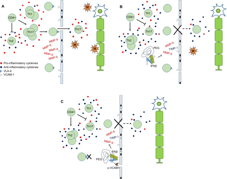
Figure 1.
Schematic depicting the role of PEG-IFNβ therapy in MS.
Notes: (A) In MS, pro-inflammatory cytokines stimulate CD4+ cells to proliferate and differentiate into Th1 and Th17 effector cells. Activated T cells express VLA-4, which interacts with VCAM-1 on endothelial cells, to facilitate crossing the BBB. In the CNS, auto-reactive T cells and macrophages result in damage to the myelin sheath, axons and neurons. Inflammatory demyelinating lesions result in the clinical presentation of MS. (B) IFNβ is conjugated to PEG to increase the molecule’s serum concentration and half-life. Proposed actions of IFNβ include modulating cytokine milieu to favor anti-inflammatory pathways, which inhibits expansion of Th1/Th17 and promotes expansion of Th2 cells. Down-regulation of VLA-4 and inhibition of MMP-9 reduce migration of activated T cells into CNS. (C) Linking of anti-VCAM-1 antibodies to the PEG tail may enhance IFNβ anti-inflammatory actions by 1) blocking interaction of leukocytes expressing VLA-4 with VCAM-1 and 2) increasing local concentration of PEG-IFNβ at BBB.
Abbreviations: BBB, blood–brain barrier; CNS, central nervous system; IFNβ, interferon beta; M, macrophage; MMP, matrix metalloproteinase; MS, multiple sclerosis; PEG, polyethylene glycol; PEG-IFNβ, pegylated interferon β; TIMP-1, tissue inhibitor of metalloproteinase-1; VCAM-1, vacular cell adhesion molecule 1; VLA-4, very late activation antigen-4.

