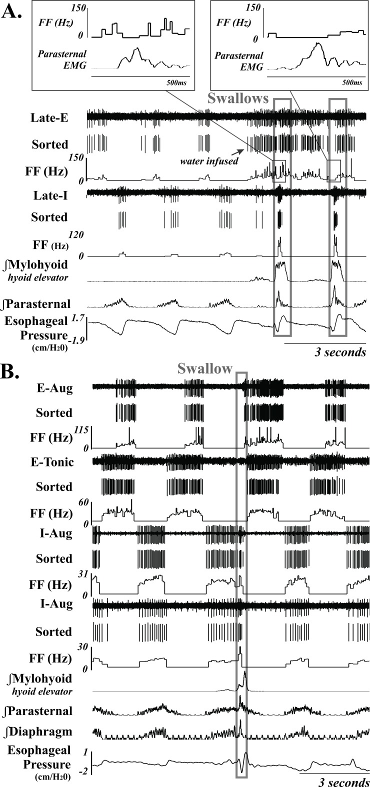
Fig 2. Example of 6 MRF neurons (3-I and 3-E) during breathing and swallow.
Unsorted and sorted spike trains, and instantaneous firing frequency (FF) (Hz) are displayed; demonstrating an increase in FF during swallow for the two I neurons. A also demonstrates more complicated swallow-related changes in the Late-E neurons firing frequency, with the second example having a longer suppression duration. B demonstrates a representative example of suppression of an E neuron during swallow. They often fire across the entire E duration, except during the execution of swallow. The swallows are outlined in a gray box. See Table 2 for anatomical location and Table 1 for neuron discharge pattern definitions. Recordings of EMG moving averages from the mylohyoid, parasternal, diaphragm (B only) are also shown.

