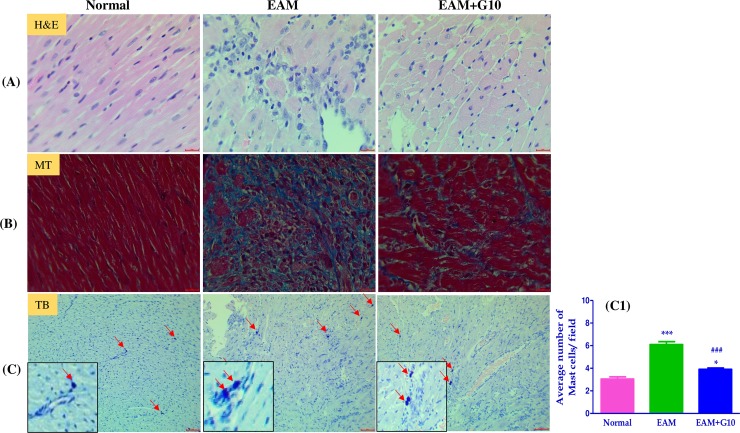
Fig 3. Effect of gallein on myocardial histological changes.
(A) H&E staining of left ventricular sections depicting infiltration of inflammatory cells, interstitial edema, vacuolization, and degeneration of cardiac fibers. (B) Masson’s trichrome (MT) staining for fibrosis (blue area) in the cross sectional tissue sections of left ventricle. (C-C1) Toluidine blue (TB) staining for mast cells of the cross sectional slices of heart and their quantification. Scale bar = 20 μm. Each bar represents mean ± SEM, n = 4–5. Normal, age matched normal rats; EAM, rats with experimental autoimmune myocarditis treated with vehicle; EAM+G10, rats with EAM treated with gallein (10 mg/kg/day). ***p<0.001 and *p<0.05 vs Normal; ###p<0.001 vs EAM.

