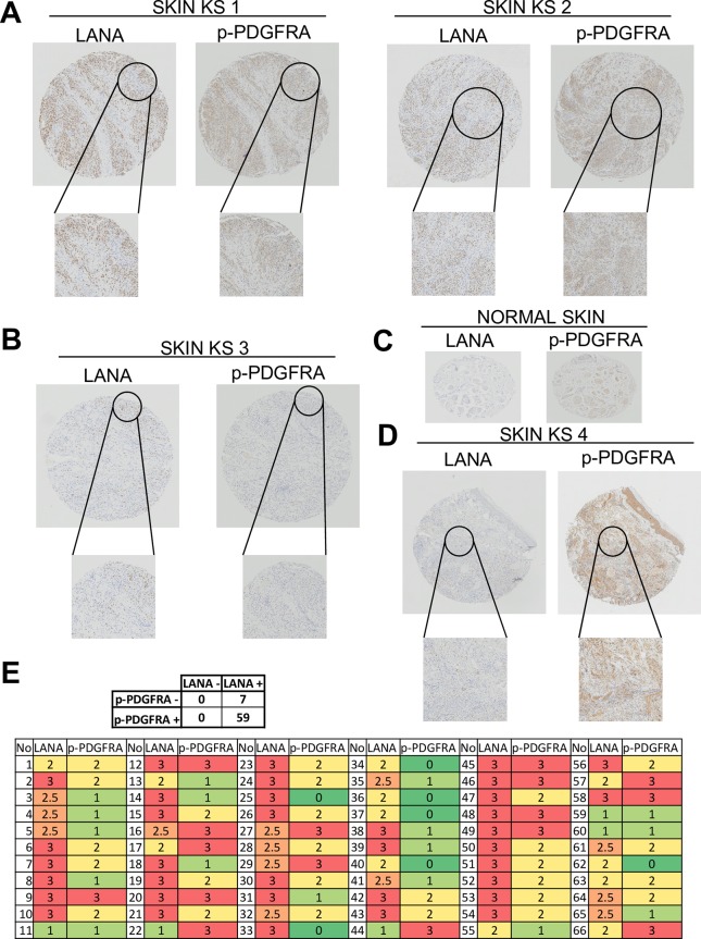
Fig 9. PDGDRA phosphorylation is consistently found in spindle-cells of AIDS-Kaposi’s sarcoma lesions and localizes to areas of KSHV infection.
(A) Staining AIDS-KS biopsies from a ACSR tissue microarray (TMA) showing that phospho-PDGFRA localizes to areas of LANA staining in two characteristic samples of the 59 out of 66 skin KS tumors which were strongly phospho-PDGFRA+ve/ LANA+ve. (B) Example of one of the 7 phospho-PDGFRA-ve (LANA+ve) AIDS-KS tumors of the TMA. (C) Normal control tissue from the TMA (skin). (D) Example of a KS tumor with strong phospho-PDGFRA staining and a low percentage of LANA+ve cells. (E) LANA and phospho-PDGFRA were scored from 0 to 3 depending on the signal strength of the antibody staining (bottom table) and total number of PDGFRA+ve/LANA+ve (59) and PDGFRA-ve/LANA+ve (7) biopsies are shown over the 66 skin AIDS-KS biopsies analyzed (top table).

