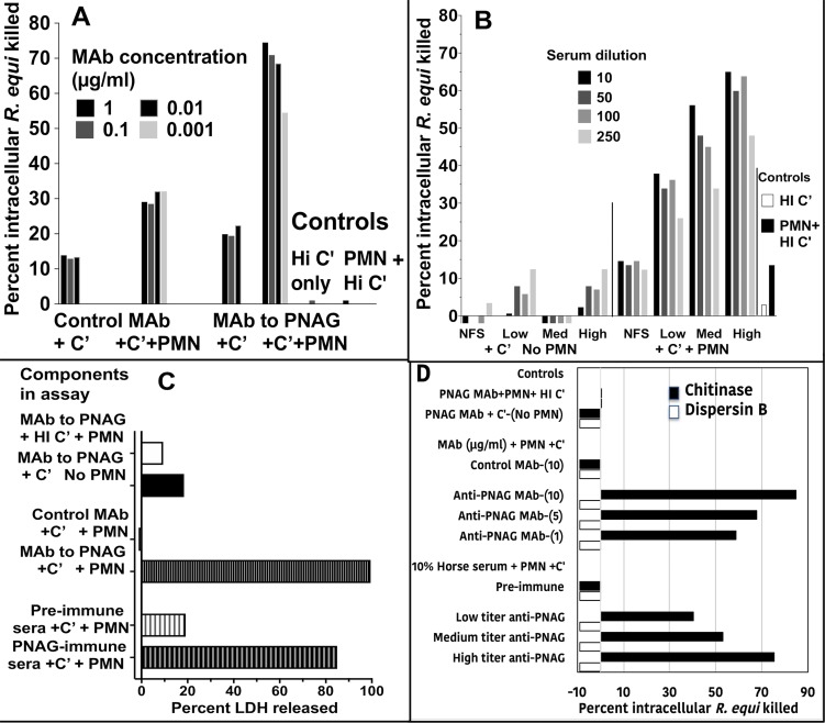
Fig 4. Opsonic killing of intracellular R. equi.
A: Maximal killing of intracellular R. equi mediated by MAb to PNAG requires both complement (C’) and PMN (C’+PMN). Background killing <5% is achieved with heat-inactivated C’ (HI C’) or PMN + HI C’. B: Pre-immune, normal foal sera (NFS) or representative immune foal sera with low, medium (Med) or high titers to PNAG obtained on the day of challenge with R. equi mediate killing of intracellular R. equi along with C’ and PMN. C: Measurement of percent cytotoxicity by LDH release shows MAb to PNAG or PNAG-immune sera plus C’ and PMN mediate lysis of infected cells. D: Opsonic killing of intracellular R. equi requires recognition of cell surface PNAG. Treatment of infected macrophage cultures with dispersin B to digest surface PNAG eliminates killing whereas treatment with the control enzyme, chitinase, has no effect on opsonic killing. Bars represent means of 4–6 technical replicates. Depicted data are representative of 2–3 independent experiments. Bars showing <0% kill represent data wherein the cfu counts were greater than the control of PNAG MAb + PMN + HI C’.

