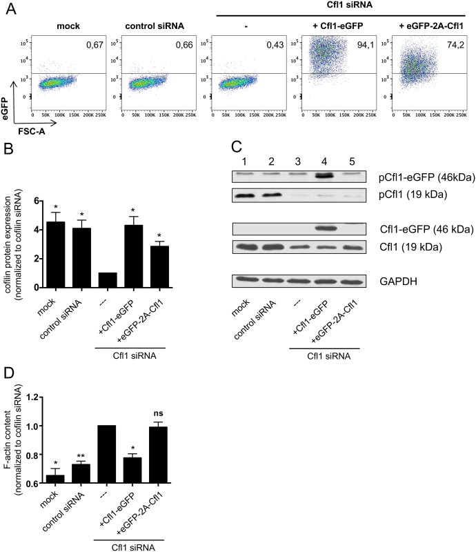
Fig 1. Cofilin expressed from an eGFP-2A-Cfl1 cassette is not functional in T-cells.
Jurkat T cells were either transfected with no siRNA (mock), a nontargeting control siRNA or a cofilin-specific siRNA (binding to the 3′ UTR), in order to downregulate endogenous cofilin. Some of the cells that received cofilin siRNA were cotransfected with Cfl1-eGFP control vector or a vector carrying the eGFP-2A-Cfl1 sequence under the control of the CMV promoter. Cells were harvested and analyzed 48 h after transfection. (A) Exemplary flow cytometric analysis of eGFP expression of transfected Jurkat cells (n = 4 independent experiments). (B) Western blot analysis of total cell lysates by staining for total cofilin and GAPDH. All samples were normalized to cells transfected with cofilin siRNA, which was set as 1. Data is represented as mean ± SEM (n = 4 independent experiments). (C) Exemplary western blot showing pCfl1, total Cfl1, and control GAPDH staining. Endogenous cofilin has a size of 19 kDa (lanes 1–3 and 5), whereas cofilin derived from the Cfl1-eGFP control vector is expressed as eGFP fusion protein (size: 46 kDa, lane 4). (D) Total cellular F-actin content was analyzed by flow cytometric measurement of phalloidin binding. All samples were normalized to cells transfected with cofilin siRNA. Data is represented as mean ± SEM (n = 3 independent experiments). Significances were calculated against Jurkat cells transfected with siRNA only. ** p < 0.01; * p < 0.05. Underlying data can be found in S1 Data. Cfl1, cofilin-1; CMV, cytomegalovirus; GAPDH, glyceraldehyde-3-phosphate dehydrogenase; ns, not significant.

