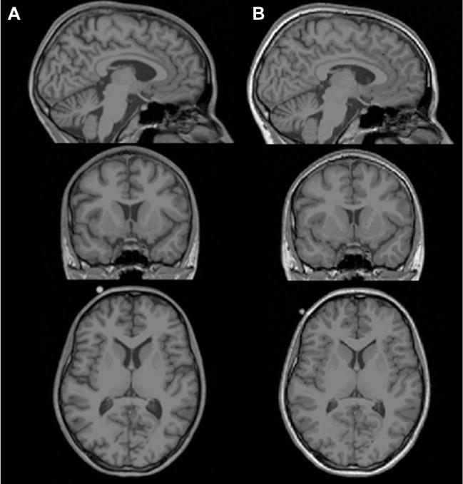
Figure 1.

Rapid macrostructural brain changes in anorexia nervosa during weight restoration. Selected sagittal (top panel), coronal (middle panel) and axial (bottom panel) T1-weighted magnetic resonance images of (A) an acutely underweight adolescent patient with anorexia nervosa at admission to an inpatient eating disorder treatment (age, 15.6 years; body mass index, 16.2) and (B) the same patient 14 weeks later at discharge following weight restoration therapy (body mass index, 19.5). The images demonstrate widespread sulcal enlargement and marked ventricle dilation in illness and rapid normalization following nutritional rehabilitation. To illustrate the dynamic alteration in brain structure in anorexia nervosa objectively, this patient was chosen from the longitudinal sample from Bernardoni et al. (20) based on her standardized body mass index change score between admission and discharge, which was equal to the sample mean. Note that changes apparent in single-subject raw magnetic resonance images may not be representative of changes detected in group analyses of processed images, which may include regionally increased brain mass both in the underweight stage and after long-term recovery (22,49).
