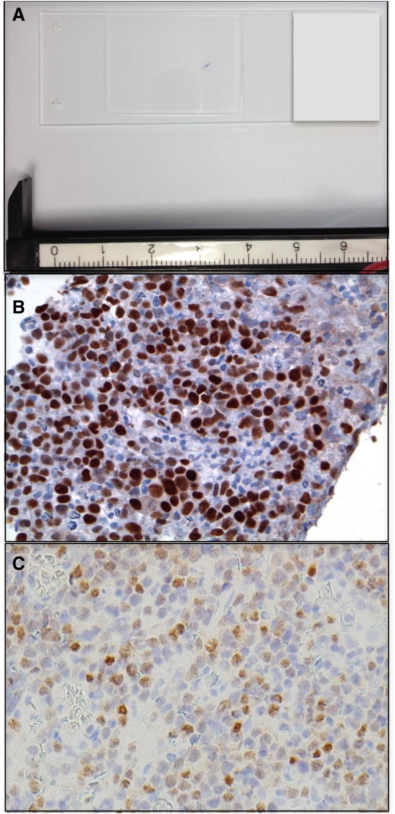
Figure 1.
PCNSL at diagnosis. (A) Example of a stereotactic brain biopsy used to diagnose DLBCL involving the corpus callosum in PCNSL. The diagnostic specimen is <2 mm in length. (B) Example of MYC protein expression, detected by immunohistochemistry in a diagnostic PCNSL brain biopsy (with diaminobenzidine detection). High expression of MYC is detected with increased frequency in PCNSL (∼50% of cases) compared with systemic DLBCL (original magnification ×40). (C) IRF-4/MUM-1 is expressed in >90% of PCNSL (original magnification ×40).

