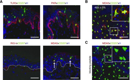
Figure 3.
LCs express MDA5 but not TLR3, RIG-I, and PKR in situ. A) Immunofluorescence double staining for markers indicated was performed on cryostat sections of untreated skin. Arrows denote MDA5+CD207+ LCs; white dotted line indicates basal membrane. B) Image of untreated epidermal sheet showing MDA5+CD207+ LCs (inset). C) Application of MDA5-specific blocking peptide revealed antibody specificity. Nuclei were counterstained with Hoechst (H). Scale bars, 50 µm.

