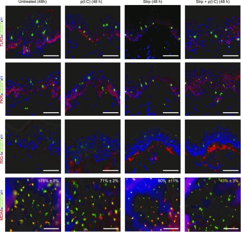
Figure 4.
LCs down-regulate MDA5 on skin barrier perturbation and p(I:C) activation on culture. Immunofluorescence double labeling of cryostat sections (TLR3, PKR, RIG-I) as well as epidermal sheets (MDA5) 48 h after culture of indicated skin explant groups. Representative merged figures with Hoechst (H) nuclear staining are shown from 3 donors in each group. Percentages in merged figures represent means ± sem of results from 3 independent experiments; MDA5+CD207+ LCs among total CD207+ LCs are shown. Tape-stripped (Strp) and p(I:C) group revealed not only less MDA5+CD207+ LCs but also much weaker MDA5 signal in LCs than respective control groups. Scale bars, 50 µm.

