Figure 5.
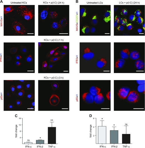
dsRNA signaling pathways are functional in LCs and KCs. Subcellular localization of MAVS, IRF3, and p65 as well as induction of IFNs and proinflammatory cytokine TNF-α in primary KCs and LCs on culture in medium or stimulation with p(I:C) was assessed at indicated time points. A, B) Distinct aggregation of MAVS as well as translocation of IRF3 and p65 from cytoplasm into nucleus was observed in stimulated KCs and LCs. Nuclear counterstain was performed with Hoechst. Localization was examined with confocal microscopy. Shown is single representative staining of 3 independent experiments. Scale bars, 5 µm. C, D) Fold change of IFN-α, IFN-β, and TNF-α mRNA expression of primary KCs (C) and sorted LCs (D) cultured for 24 h without or with p(I:C) is shown. Results expressed as means ± sem are from 6 independent experiments with 6 different donors. *P < 0.05, **P < 0.01 compared to unstimulated cultured group.

