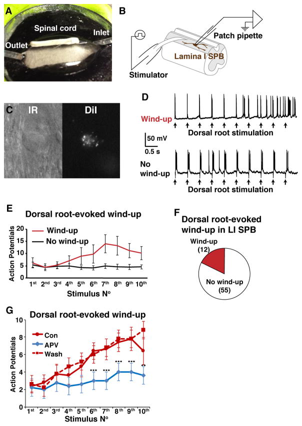
Figure 1. Dorsal root-stimulation induces wind-up in 18% of lamina I SPB neurons.
A–B. Photograph (A) and schematic (B) of recording set up in whole spinal cord preparation. Whole-cell patch clamp recording was made from the lamina I SPB neurons. C. Infrared (IR) and fluorescent image of a lamina I SPB neuron that is labeled with DiI. D. Example traces of wind-up and no wind-up in response to 2 Hz dorsal root stimulation. E. Wind-up is observed in a subset of lamina I SPB neurons in response to 2 Hz root stimulation. F. Pie chart illustrating fraction of lamina I SPB neurons that show wind-up. G. Treatment with the NMDA antagonist APV (50 μM) significantly reduced wind-up in lamina I SPB neurons in response to 2 Hz root stimulation, which recovered upon wash. Data are mean ± SEM (n = 5 cells, paired; asterisks indicate significantly different than control, ** p < 0.01, *** p < 0.001, Two-way ANOVA followed by Dunnett’s multiple comparison test).

