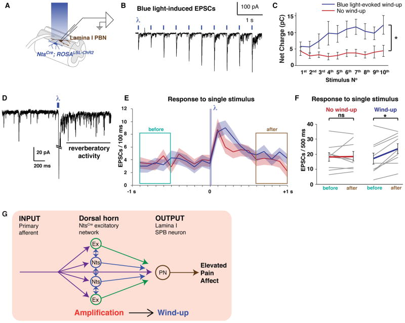
Figure 5. NtsCre neurons form an extensive excitatory network.
A – C. Whole-cell patch clamp recording from lamina I SPB neurons upon optogenetic stimulation of NtsCre neurons (0.1 Hz ×10, 5 ms duration). Example traces from recorded cells that received no input (A), monosynaptic and polysynaptic input (B), or polysynaptic input alone (C) from NtsCre neurons. D. Summary data; n = 30 cells. E – G. Whole-cell patch clamp recording from NtsCre neurons upon optogenetic stimulation of NtsCre neurons (0.1 Hz ×10, 5 ms duration). Example traces from recorded cells that received no input (E) (inward current is due to opening of ChR2 in the recorded cell), monosynaptic and polysynaptic input, as indicated by the blue arrow (F), or polysynaptic input alone, as indicated by the green arrows (G) from NtsCre neurons. H. Summary data; n = 25 cells.

