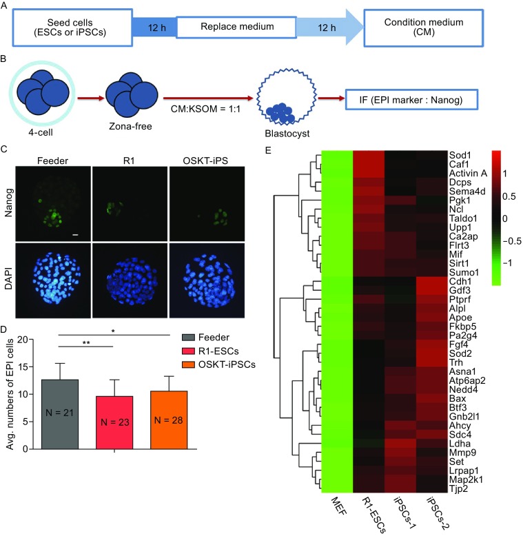
Figure 2.

Secretions from ESCs and iPSCs affect EPI development. (A) Schematic of the method used to collect the condition medium. (B) Experimental design. Zona-free embryos at 4-cell stage were treated in the mixed medium containing KSOM and CM and then immunostained at E4.5 to test the effect of the condition medium on early embryo development fate. CM, condition medium. (C) Nanog immunostaining in E4.5 embryos treated with condition medium from feeder, R1 ESCs and iPSCs. Nuclei were stained with DAPI (Blue). Scale bars, 20 μm. (D) Average numbers of EPI cells (Nanog-positive cells) in condition medium-treated embryos at E4.5. Error bars indicate SD. *P < 0.05; **P < 0.01 by ANOVA. N is the number of embryos examined. (E) Heatmap of ESC and iPSC-secreted proteins at high expression levels. The heatmap was plotted with relative protein expression
