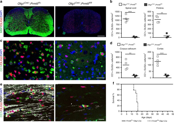
Fig. 2.
Genetic ablation of Prmt5 in OPCs induces severe hypomyelination and impaired OPC differentiation. Confocal images of P14 spinal cord (a) and corpus callosum (c, e) sections from controls (Olig1Cre/+;Prmt5fl/+) and Prmt5 mutants (Olig1Cre/+;Prmt5fl/fl) stained for MBP (a: green, scale bar = 200 μm; e: white, scale bar = 20 μm), CC1 (c, e: green, scale bar = 20 μm), OLIG2 (a: red, scale bar: 200 μm; c, e: red, scale bar = 20 μm). DAPI (blue) as nuclear counterstain. (b, d) Scatter plot represents the average number of CC1+/OLIG2+ cells quantified in spinal cord and fimbria (b) and in corpus callosum and cortex (d) of four control mice (Olig1Cre/+;Prmt5fl/+) and three Prmt5 mutants (Olig1Cre/+;Prmt5fl/fl). Student’s t test, **p < 0.01, ***p < 0.001. f Survival curve showing comparison in controls (Olig1Cre/+;Prmt5fl/+) and nine Prmt5 mutants (Olig1Cre/+;Prmt5fl/fl)

