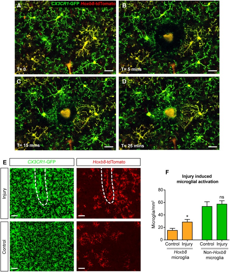
Fig. 7.
Hoxb8 microglia have similar response to damage as non-Hoxb8 microglia. (A-D) Stills from a time-lapse movie of microglial response to damage induced by focused laser ablation in brain cortex, using in vivo multiphoton imaging. Scale bars: 30 μm. (A) Hoxb8 (yellow) and non-Hoxb8 (green) microglia in their resting state before injury. A video of the acute response is provided (Movie 1). (B-D) Successive time points as the microglial processes converge towards the site of injury. (E) Microglial activation induced by needle-poke injury (white dashed lines) in the mouse brain cortex observed 7 days postinjury. Scale bars: 50 μm. (F) Hoxb8 microglia show a 1.9-fold increase in numbers around the injury site compared with a 1.1-fold increase in non-Hoxb8 microglia. n=5 mice, ns, non-significant; *P<0.05; data are mean±s.e.m. See also Fig. S7 and Movie 1.

