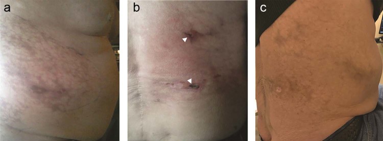
Fig. 2.
(A) Right flank mottling immediately after right inferior phrenic artery particle embolization. (B) Photograph of the right flank 3 weeks postembolization. Note two small 1-cm eschars from focal necrosis and ulceration (arrowheads). (C) Photograph of the right flank approximately 3 months later showing healing of the ulcerated areas with residual skin pigmentation.

