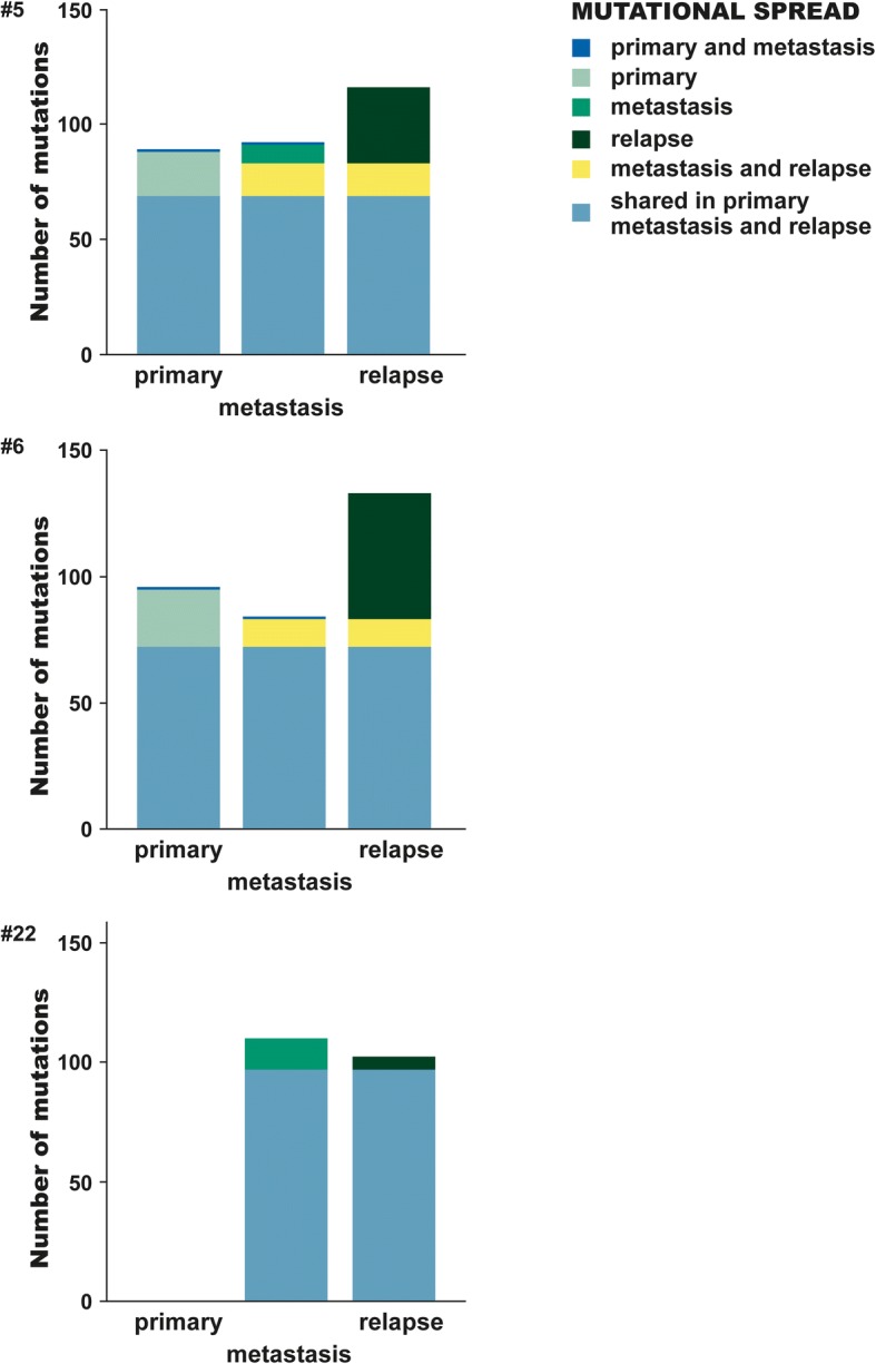
Fig. 6.

Mutations located in the primary tumour, synchronous liver metastasis and metachronous liver metastasis. Patient(#) 5 and 6 had all three sites successful sequenced and analysed, #22 had a primary tumour with insufficient residual tumour content and therefore not analysed. The bar of each site (primary tumour, synchronous metastasis, relapse) are coloured according to the identified variants being shared with another site or private
