
Figure 5.
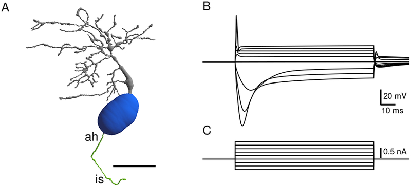
Excitability of reconstructed bushy neuron. A. Reconstruction of a fluorescently labeled mouse bushy cell from the data set of Campagnola and Manis, (2014). The cell was filled with the fluorescent dye AlexaFluor 488, and reconstructed from a multiphoton image stack using Neuromanitc (Myatt et al., 2012). The cell was then decorated with channels as indicated in Table 2. Grey: Dendrites, blue: soma, green: axon hillock (ah) and initial segment (is). Scale bar: 20 microns. B. Traces in response to current injections at the soma, showing canonical bushy cell response (parameters of channel decoration are shown in Table 1). C. Current steps for panel B.