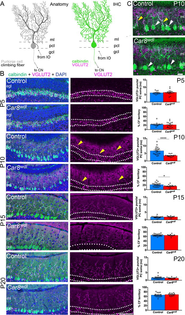
Fig. 1.

(a) Schematics of the olivo-cerebellar-nuclei circuit (left) and CF terminals on a PC (right). Abbreviations: IHC = immunohistochemistry, ml = molecular layer, pcl = Purkinje cell layer (white dotted lines), gcl = granule cell layer, cn = cerebellar nuclei, IO = inferior olive. (b) VGLUT2 and calbindin double staining reveals premature CF elimination and delayed CF translocation into the molecular layer at P10 (scale bar = 50 μm). The number (no) of VGLUT2-positive puncta on PC somata (p<0.0001) and percent CF territory (p=0.0159) were significantly different between Car8wdl and control mice at P10. In the graphs, * = 0.0159, and **** < 0.0001. Error bars reflect ± the standard of the mean (SEM). Yellow arrowheads point to the CF terminals that have translocated from the PC somata onto the PC dendrites. (c) Higher magnification of VGLUT2-positive CF terminals on PC somata (white arrowheads) and dendrites (yellow arrowheads) at P10 (scale bar = 20 μm). Control mice have more VGLUT2-positive CF terminals on PC somata and dendrites compared to the Car8wdl mutant mice.
