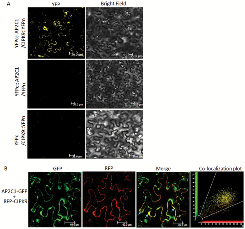
Fig. 3.
Interaction analysis of AP2C1 and CIPK9 by bimolecular fluorescence complementation (BiFC) and co-localization assays. (A) Nicotiana benthamiana cells co-infiltrated with YFPc::AP2C1/CIPK9::YFPn showing reconstitution of the yellow fluorescent protein (YFP) signal in the cytosol, while co-infiltration of YFPc::AP2C1/YFPn and YFPc/CIPK9::YFPn (negative controls) shows no YFP fluorescence, Scale bars are 20 µm. (B) N. benthamiana epidermal cells co-transformed with RFP-CIPK9 and AP2C1-GFP showing merger of the two signals in the cytosol as yellow fluorescence. The scatter-plot showing maximum yellow dots in the common region confirms the co-localization of the two proteins. Scale bars are 40 µm.

