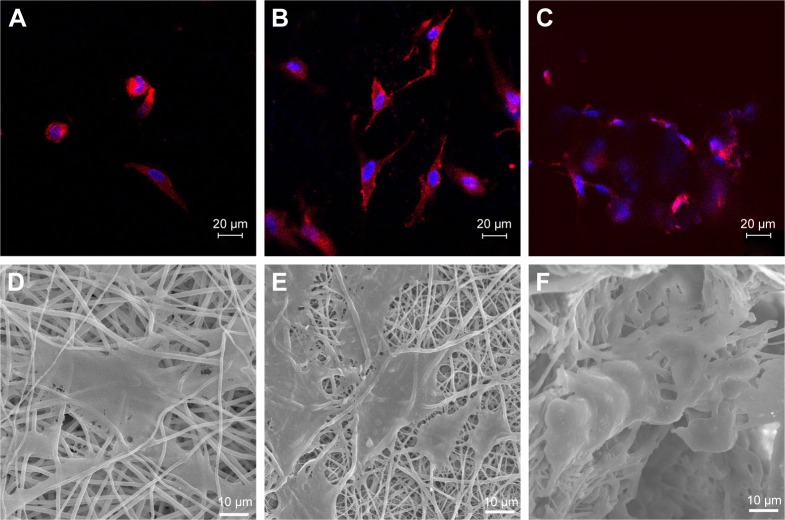
Figure 5.
Morphology of rADSCs on scaffolds.
Notes: Representative CLSM images of rADSCs on scaffolds after 1 day: PP (A), PP–B (B), 3D (C), cytoskeleton (red), and cell nuclei (blue). Representative SEM images of ADSCs on scaffolds after 7 days: PP (D), PP–B (E), and 3D (F).
Abbreviations: 3D, three dimension; CLSM, confocal laser scanning microscopy; PP, poly(lactide-co-glycolide)/polycaprolactone; PP–B, PP–bone morphogenetic protein-2; rADSCs, rat adipose-derived stem cells; SEM, scanning electron microscopy.

