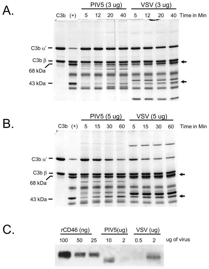
Figure 4.
Comparison of virion-associated CD46 cofactor activity from purified PIV5-CD46 versus VSV-CD46. A and B) C3b cleavage assays were reconstituted in vitro using purified C3b, Factor I and either 3 ug (panel A) or 5 ug (panel B) of purified PIV5 and VSV derived from CD46-expressing CHO cells. After the indicated times at 37°C, samples were analyzed by SDS-PAGE and coomassie blue staining. The positions of the α′ and β chains of C3b are indicated, and arrows show the position of the 68 and 43 kDa cleavage products. C) Levels of PIV5- and VSV-associated CD46 from purified virus preparations were analyzed by western blotting along with the indicated amounts of purified rCD46.

