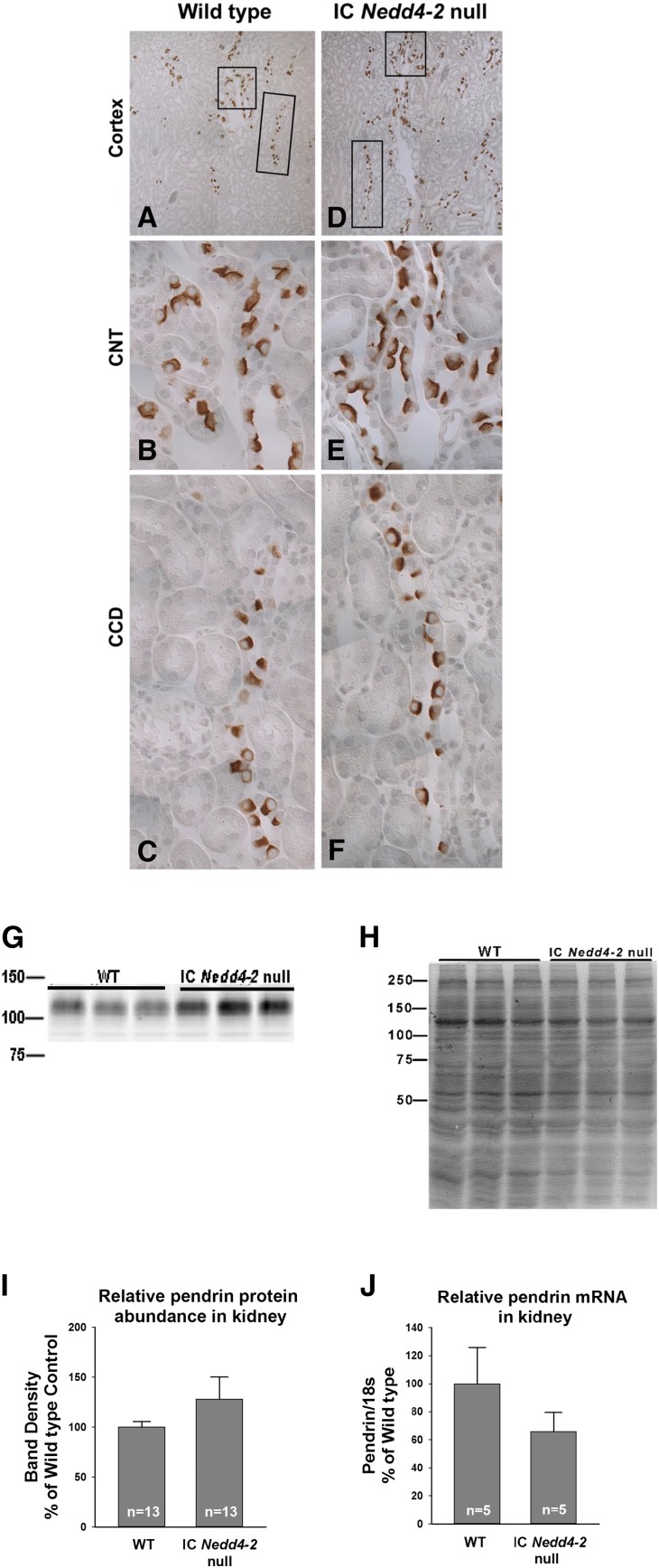
Figure 9.
Pendrin protein abundance is not significantly changed with intercalated cell (IC) Nedd4–2 gene ablation. Cortical sections from a representative IC Nedd4–2 knockout mouse and a wild-type (WT) littermate were labeled for pendrin. A and D show pendrin labeling at low magnification. Insets show typical (B and E) connecting tubules (CNTs) and typical (C and F) cortical collecting ducts (CCDs) at higher magnification. G shows a representative immunoblot of kidney lysates from IC Nedd4–2 knockout mice and WT littermates probed for pendrin and its respective Coomassie blue gel, which (H) confirms protein loading. I shows that pendrin band density is similar in kidney lysates from IC Nedd4–2 null mice and WT littermates. J shows that kidney pendrin (Slc26a4) mRNA when normalized to 18S mRNA is the same or reduced with IC Nedd4–2 gene ablation.

