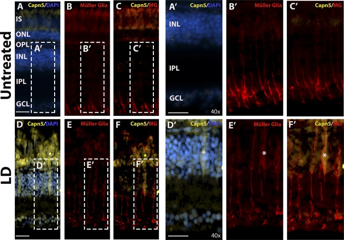
Figure 7.
capn5a is expressed in the Müller glia in response to acute LD. (A–C′) IHC for Capn5 expression (A) and the Müller glial marker Zrf–1 (B) in the undamaged (UT) adult retina. No colocalization of Capn5 and Müller cells is observed. (A′–C′) show an enlarged image of the boxed regions in (A–C). (D–F) IHC for Capn5 and Müller glia in the LD retina. Capn5 expression is induced in the INL, where it colocalizes with reactive Müller glia (asterisk). (D′–F′) show enlarged images of the boxed regions in (E–F). IPL, inner plexiform layer. Scale bars: 100 μm in (A–F) and 500 μm in (A′–F′).

