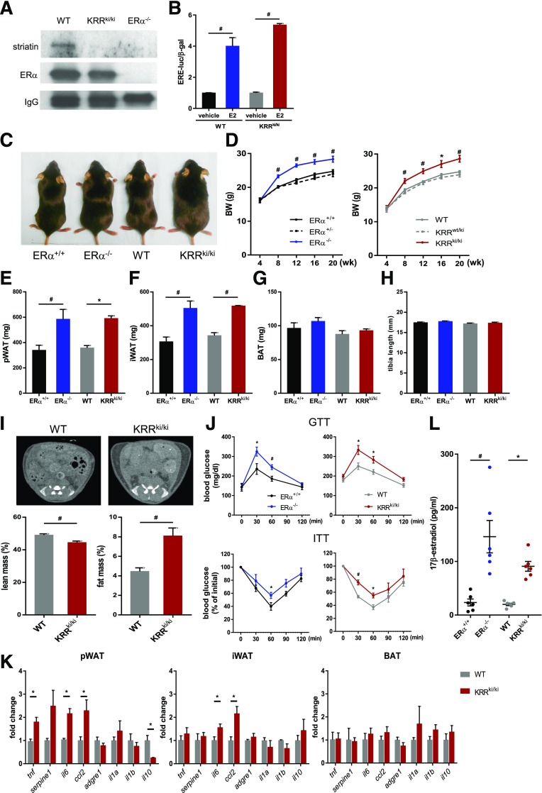
Figure 1.
Membrane-initiated signaling is essential for ERα-mediated metabolic homeostasis. A: Coimmunoprecipitation of ERα with striatin. Proteins were extracted from uterus tissue of WT, KRRki/ki, and ERα−/− mice, immunoprecipitated using an ERα antibody, and immunoblotted using an antibody against striatin. A representative immunoblot is shown. B: Carotid artery VSMCs from WT and KRRki/ki mice were transiently cotransfected with an estrogen response element–driven luciferase reporter plasmid and a β-galactosidase expression plasmid. Cells were treated with vehicle or E2 for 24 h (n = 3). #P < 0.01. C: Gross appearance of ERα+/+, ERα−/−, WT, and KRRki/ki mice. D: Body weight (BW) of female mice (n = 8–12) over the course of the study. *P < 0.05, #P < 0.01 vs. ERα−/− (left panel) or WT (right panel) mice. E–H: Weights of pWAT and iWAT and BAT fat pads as well as the tibia lengths of WT, KRRki/ki, and ERα−/− mice at 12 weeks of age (n = 8–12). *P < 0.05, #P < 0.01. I: Evaluation of fat and lean mass via CT imaging (n = 9–11). #P < 0.05. J: Glucose tolerance test (GTT) and insulin tolerance test (ITT) results were assessed (n = 6–8). *P < 0.05, #P < 0.01. K: qRT-PCR analysis for inflammatory cytokines (n = 3–6 per group). *P < 0.05. L: Serum E2 levels (n = 6 per genotype). *P < 0.05, #P < 0.01. Data are represented as mean ± SEM.

