
Fig. 1. Schematic representation of noncovalent imprinting process and corresponding surface characterizations by microscopy.
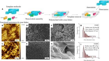
(A) Molecular memory is introduced on the sensor surface by copolymerizing functional monomer and cross-linker in the presence of the analyte, which acts as a molecular template. After elution of the analyte, complementary binding sites are revealed complimentary in size and shape to template by creating molecular memory on the surface that allows specific rebinding of the target molecule. The recognition sites obtained in this manner have binding affinities approaching those demonstrated by antibody-antigen systems. Tapping-mode atomic force microscopy (AFM) analysis of cortisol-selective polymer (B) and its corresponding control (C). Scanning electron microscopy (SEM) images of cortisol-selective polymer (D and F) and its control (E and G) with two different magnifications. Pore size distribution for cortisol-imprinted (H) and nonimprinted polymers (NIPs) (I). The BJH method was applied to calculate pore size distribution from experimental isotherms using the Kelvin model of pore filling. The method applies only to the mesopore and small macropore size range.