
Fig. 2. Schematic representation of membrane integration and device characteristics.
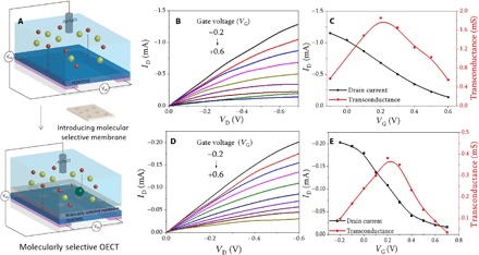
(A) Schematic drawing of integration of MSM to OECT device consisting of PEDOT:PSS channel coated on an indium tin oxide glass substrate with the cortisol-selective membrane separating the PEDOT:PSS channel from the electrolyte solution, gated with an Ag/AgCl electrode. Device characteristics demonstrated by output and transfer curves before (B and C) and after (D and E) membrane integration. The output and transfer curve measurements were conducted in artificial sweat solutions.