
Fig. 3. Testing of MS-OECT device for ex situ analysis.
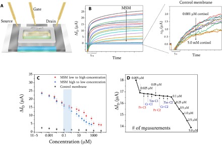
(A) Schematic drawings of planar-gated MS-OECT device on the SEBS substrate for drain current measurements. PDMS, polydimethylsiloxane. Ex situ measurement for MSMs (inset: control membrane) (B) and their corresponding calibration curves (the calibration curve for high-to-low concentration is based on fig. S6B, and the highlighted area shows the physiological range of cortisol in human sweat) (C). The measurements were conducted in artificial sweat with increasing concentrations of cortisol. The selectivity of the MS-OECT device was evaluated in the presence of structural analogs of cortisol including progesterone, cortisone, and testosterone in artificial sweat. The measurement was started with 0.005 mM cortisol, and it was gradually increased up to 5.0 μM. The concentrations of interferent were increased step-wise for progesterone (Pr-C1, 0.025 μM; Pr-C2, 0.5 μM), cortisone (Cr-C1, 0.025 μM; Cr-C2, 0.5 μM), and testosterone (Tes-C1, 0.025 μM; Tes-C2, 0.5 μM) to ensure that they do not involve a binding process (D). The drain current measurement of each run is shown in fig. S25.