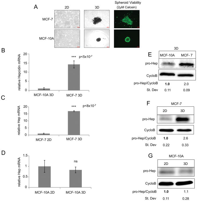
Figure 2. Hepcidin is increased in MCF-7 breast cancer spheroids compared to non-tumor spheroids and is induced from 2D to 3D culture of breast cancer cells.
(A) Phase-contrast imaging of MCF-7 and MCF-10A cells grown in 2D and 3D and fluorescent imaging of spheroids stained with 2μM calcein-AM. (B–D) RT-qPCR of hepcidin mRNA (normalized to cyclophilin A) in MCF-7 and MCF-10A cells grown in 2D and 3D. (E–G) Western blot of pro-hepcidin using Cyclophilin B as internal control. Scale bar: 10μm (2D), 200μm (3D)

