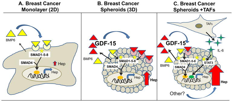
Figure 9. Working model of regulation of hepcidin in breast cancer spheroids.
Hepcidin expression is increased in breast cancer cells relative to non-cancer cells. A. In MCF-7 cells grown as 2D monolayers, BMP6 plays a dominant role in the cancer-dependent increase in hepcidin through activation of a SMAD1-5-8 signaling pathway. B. In breast cancer spheroids, spatial control of hepcidin synthesis is exerted by GDF-15, which augments SMAD 1-5- 8 signaling to further increase hepcidin synthesis. C. The microenvironment is an additional source for increased hepcidin in breast cancer cells, in part due to production of IL-6 by tumor-associated fibroblasts (TAFs).

