Figure 2.
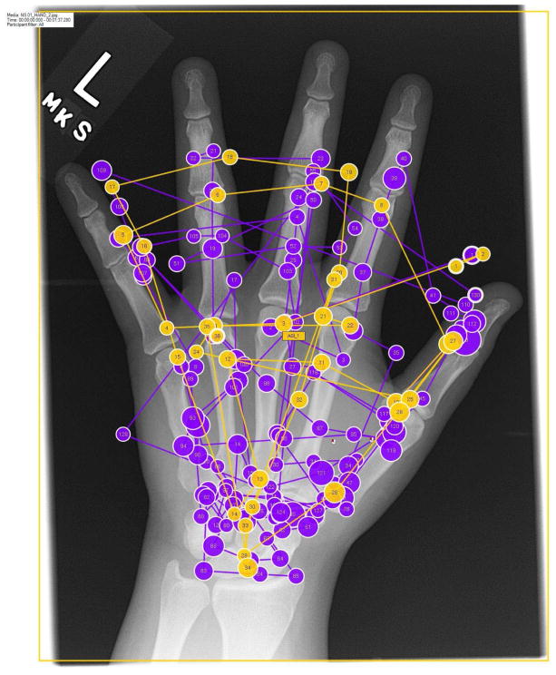
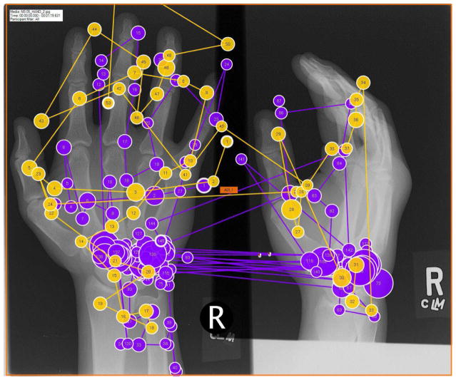
Visual search patterns of radiologists captured by eye-tracking technology near the end of a normal work-day (4 PM during a 9hr 8AM–5PM shift, “Not Fatigued” Session) vs following an overnight shift (8 AM following a 9hr 10PM–7AM shift, “Fatigued” Session). A) Example of one of the normal test images with the search pattern of a single reader in fatigued (purple) and non-fatigued (yellow) states superimposed. Each circle represents a gaze fixation (location where foveal vision is directed). The size of the circle represents fixation dwell time with larger circles reflecting increased dwell times. The lines between the fixations represent saccades, which are rapid eye movements between fixations. Notice the significant increase in gaze fixations in the fatigued state. B) Example of one of the abnormal test images (carpal fracture, which is obscured by the eye-tracking pattern). Notice the increase in gaze fixations and dwell time in the carpus during the fatigued state.


