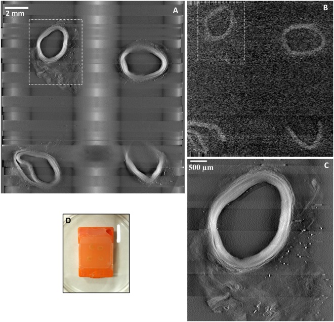
Figure 1.
Comparing a prototype tomographic scanner dedicated to histopathology (Tomopath) with a commercial tabletop micro-CT scanner. (A) Cross-sectional image at 1.3 mm level below the surface of a paraffin block from the Tomopath scanner. Left-anterior-descending (LAD) coronary artery segments of a deceased HIV patient are embedded in the block. The vessel walls appear bright against a dark background from the x-ray absorption contrast between tissue and paraffin medium. The grid-like shadows are cast by the plastic grid of the embedding cassette. (B) Cross-sectional image at the same level from the commercial tabletop micro-CT scanner. (C) Zoomed-in view of the top left LAD segment outlined in A from the Tomopath scanner. The layers of the vessel wall are visible. The scan times of the micro-CT and the Tomopath scanners were 2.75 hours and 15 minutes, respectively. (D) Picture of the intact paraffin block prior to histological sectioning.

