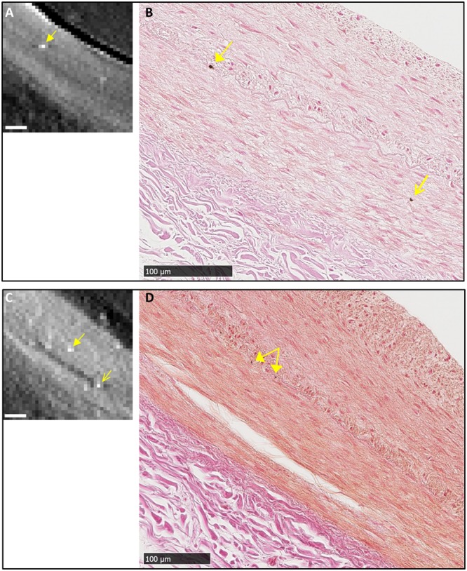
Figure 5.

Two instances of un-correlated finds of micro calcifications in the media layer. (A) and (B) Near an IEL micro calcification visible in both tomography and histology (solid arrows), an isolated micro calcification in the media layer is seen in the histological image of panel B (open arrow), but not seen in the tomographic image of panel A. (C) and (D) Near focal calcifications in the IEL seen in both the tomographic and histological images (solid arrows), a focal calcification in the media is seen in the tomographic image (open arrow in panel C) but absent in the histological image panel D. Its location was determined based on a tear in the media visible in both images. All scalebars are 100 µm.
