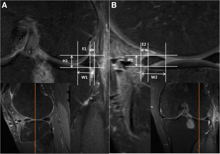
Fig. 1.
Measurements of graft extrusion or intrusion in the mid-sagittal plane and mid-coronal plane. From the mid-coronal knee MRI section (a), the radial extrusion (E1) was measured as the distance between the outer edge of the articular cartilage of the tibial plateau and the outer edge of the allograft. Also, the height (H1) and the width (W1) of the graft were measured. The radial relative percentage of extrusion (RPEr) was defined as the percentage of the width of extrusion relative to the width of the entire allograft (E1/W1 × 100). Similarly, the anterior relative percentage of extrusion (RPEa) was calculated after measuring the anterior extrusion (E2) and graft width (W2) from the mid-sagittal section (b). Graft height in sagittal section (H2) was also measured. Extrusion was expressed as a positive value and intrusion as a negative value

