Fig. 4.
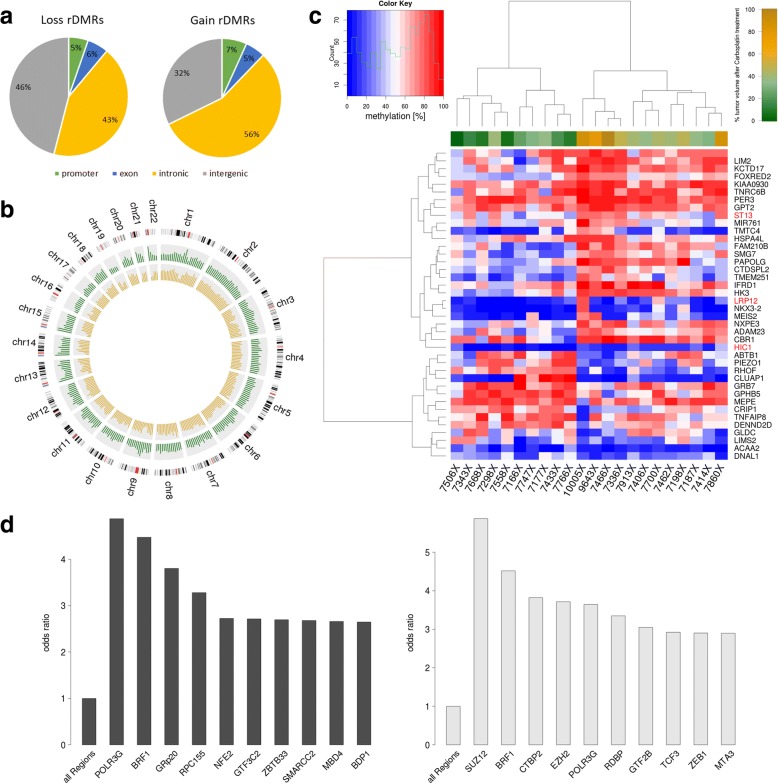
Carboplatin-resistant tumors exhibit distinct changes in DNA methylation. a Pie chart of the genomic distribution of carboplatin rDMRs (p value < 0.05). b Circos plot of the localization and frequency of the 837 most significantly differentially hypermethylated regions (p value 0.0001 and absolute correlation of methylation to relative tumor volume > 0.5). Red: hypermethylated regions in responders (263 regions); blue: hypermethylated regions in non-responders (574 regions). c Heatmap representation of unsupervised hierarchical clustering analysis of 40 candidate promoter-associated carboplatin rDMRs. The upper bar represents the sensitivity phenotype of the PDXs (red: non-responders; green: responders). d Barplot showing the most significantly differentially hyper- and hypomethylated ENCODE-defined TFBS in carboplatin non-responders. e Tumor vs normal methylation differences show methylation differences between non-responders and responders in large hypomethylated blocks (LHBs) on chromosome 1 as example. Green highlighted strong responder (relative tumor volume < 9%, n = 4), light green intermediate responder (relative tumor volume > 9%/< 30%, n = 5), gray weak responder (relative tumor volume > 30%/< 78%, n = 4), and black non-responder (relative tumor volume > 78%, n = 4). The dark red line indicates LADs (lamina-associated domains) that are associated in location with LHBs. TFBS transcription factor binding site, CGI CpG island

