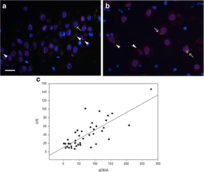
Fig. 3.
Colocalization of poly-GR and aDMA. Poly-GR neuronal cytoplasmic inclusions in the dentate fascia and CA4 of the hippocampus. Sparse poly-GR inclusions in the dentate fascia show colocalization with aDMA (arrow). Note that not all poly-GR aggregates contain aDMA (arrowheads) (a). Moderate poly-GR inclusions show colocalization with aDMA in CA4 (arrows). Again, not all poly-GR aggregates contain aDMA (arrowheads) (b). Scale bars: 10 μm. Plot shows the association of poly-GR and aDMA neuronal inclusions in the dentate fascia. The line shows linear regression (r = 0.77) (c)

