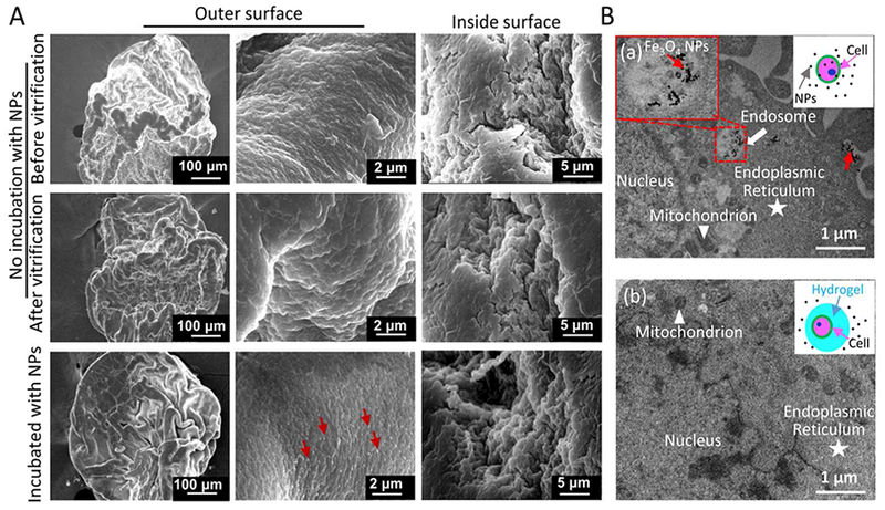
Figure 4.

SEM images of cell-alginate hydrogel constructs under different conditions and cell uptake of Fe3O4 NPs. A) No evident difference in the morphology of hydrogel microcapsules before and after vitrification with nano-warming. When the constructs (microcapsules) were incubated with Fe3O4 NPs overnight at 4 °C, the NPs appeared only on the surface of the microcapsules. The red arrow indicates Fe3O4 NPs. B) Non-encapsulated pADSCs (a) and pADSCs-hydrogel constructs (b) were incubated with Fe3O4 NPs for 10 h at 37 °C. The NPs were only found in non-encapsulated cells (a), the red arrow indicates NPs in endosomes.
