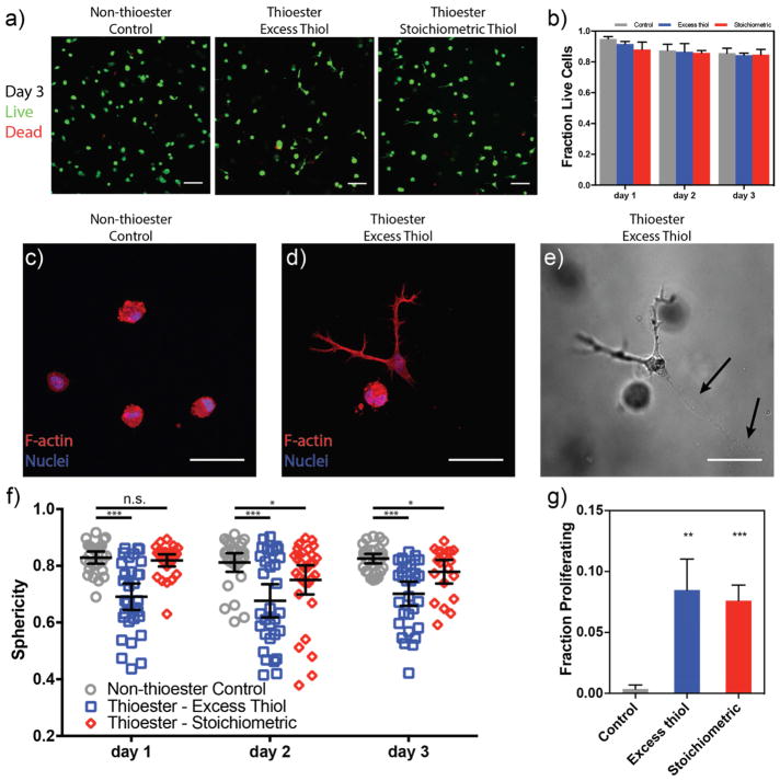
Figure 4. hMSCs encapsulated in thioester hydrogels are viable, acquire elongated morphologies and have increased proliferation relative to static networks.
a) Encapsulated cells were stained with Calcein-AM (green, live) and Ethidium homodimer-1 (red, dead) each day and visualized by confocal microscopy. Images shown at 72 h post-encapsulation. b) Quantified fraction of viable cells from confocal z-stacks. All conditions promote cell survival over three days of culture. c–d) Representative images of cell shape on day 3: cells in static networks remain rounded (c), while a subset of those in thioester networks take on a spread morphology. e) Transmitted light image of hMSC in (d) with visible cell tracks left behind (black arrows). f) Sphericity of the three-dimensional cell shapes was analyzed during culture within hydrogels. Thioester networks formed with excess thiols (blue squares) show dramatically increased spreading (i.e. lower sphericity) compared to static hydrogels within one day of encapsulation and this difference is retained throughout the experiment. Thioester networks formed with stoichiometric thiols begin to spread after one day in culture. d) hMSCs in thioester networks proliferate more than those in static hydrogels. Cells were incubated with EdU on day 2 to visualize dividing cells. Scale bars: 100 μm (a), 50 μm (c–e). Plots are shown as mean ± std. dev. (b&g), ± 95% conf. interval (f). *,**,*** indicate p < 0.05, 0.01, 0.001 respectively.

