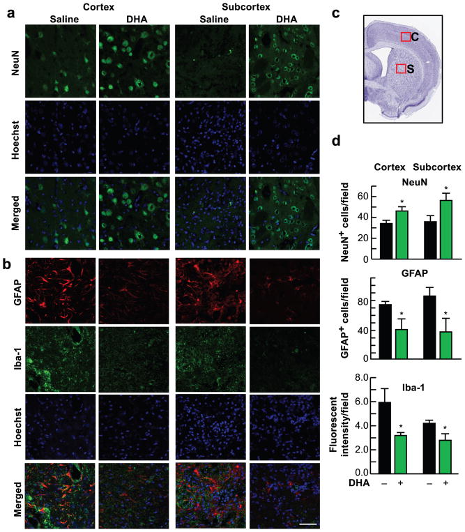
Fig. 4.
DHA attenuates cell damage 4 weeks after MCAo. (a, b) Representative images of NeuN (green), GFAP (red), Iba-1 (green) and Hoechst staining in the cortical and subcortical regions. Scale bar: 50 μm. (c) Coronal brain diagram showing locations of regions for cell count in peri-infarct cortex (C) and subcortex (S). (d) Quantification of NeuN, GFAP and Iba-1 in peri-infarct cortex and subcortex. Values are mean ± SEM; n=7–9 per group. * p <0.05 vs. saline group (Repeated measured ANOVA followed by Bonferroni test)

