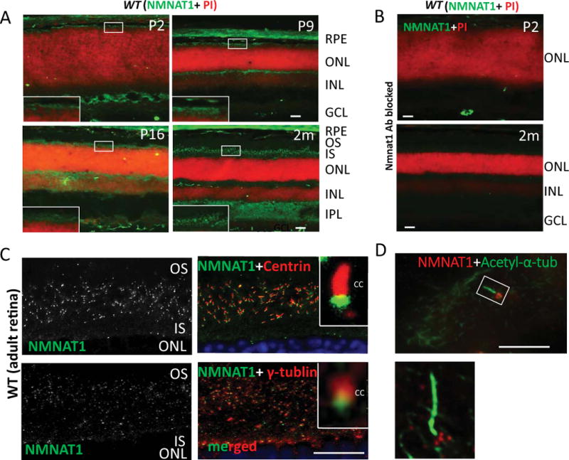
Figure 1. NMNAT1 localizes to the basal body of mouse photoreceptors.

(A) NMNAT1 expression in wild-type frozen sections is detected in the photoreceptor cell layer beginning at P2 and continues throughout adult stages (NMNAT1 shown in green; propidium iodine (PI) shown in red indicating nuclear staining). (B) Cryosections of WT retina treated with a NMNAT1 blocking peptide in combination with NMNAT1 antibody shows lack of NMNAT1 staining, confirming the specificity of the NMNAT1 antibody. (C) Cryosections of WT retina co-stained with anti-NMNAT1, pan-Centrin (an axoneme marker) and γ-tubulin (a marker of the basal body) antibodies. Inserts: high magnification images show NMNAT1 positive signals near the basal body of the photoreceptor. (D) Co-staining of ciliated hTERT-RPE1 cultured cells with NMNAT1 and acetylated α-tubulin (an axoneme marker) shows NMNAT1 positive signal near the basal body. Scale bar: A–B: 20um; C: 10um; D: 2um. RPE= retinal pigment epithelium, ONL= outer nuclear layer, INL= inner nuclear layer, IPL= inner plexiform layer, GCL= ganglion cell layer, OS= outer segment, IS= inner segment, CC= connecting cilium.
