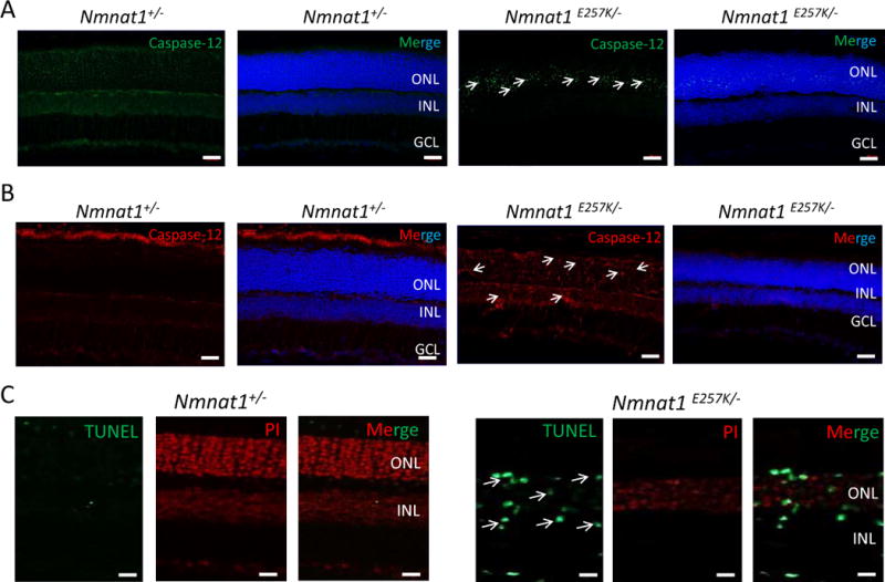
Figure 6. Light damage induces cell death in Nmnat1E257K/− retinas at 7 and 14 days after light exposure.

Cryosections of 8 weeks-old Nmnat1E257K/− retinas stained for Caspase-12, a marker of ER stress, after light exposure (A, B). Caspase-12 is not detected in the ONL of Nmnat1+/− retinas while strong staining is observed in the ONL of Nmnat1E257K/− retinas at 7 days (A) and 14 days (B) after light exposure. A subset of positive signals is marked by arrowheads. (C) TUNEL-positive cells (green) are detected mostly in the INL and ONL of paraffin embedded Nmnat1E257K/− retinas at 14 days after light exposure (right), while no signal is observed in control retina after light exposure (left). Scale bar: 20 μm. DAPI (blue) was used to counterstain nuclei in Panels A and B. Propidium Iodide (PI) was used to counterstain nuclei in Panel C. ONL= outer nuclear layer, INL= inner nuclear layer, GCL= ganglion cell layer.
