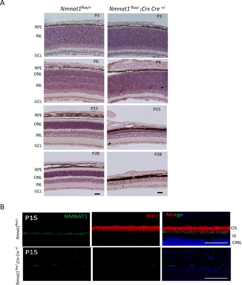
Figure 8. Conditional ablation of Nmnat1 in photoreceptors using Crx-Cre causes rapid retinal degeneration.

(A) H&E staining on Nmnat1 Crx-Cre cKO (Nmnat1Flox/−; Crx-Cre) mice reveals defects in lamination at P6 and strong loss of retinal tissue by P15. At P28, there is a significant reduction in the overall thickness of the retina as well as the photoreceptor ONL layer in Nmnat1Flox/−; Crx-Cre mutant retina compared to Nmnat1Flox/+ control retina. (B) Rhodopsin (red) staining shows complete loss of outer segments in Nmnat1Flox/−; Crx-Cre retina at P15, consistent with strong loss of NMNAT1 immunoreactivity (green). In comparison, the expression of Rho in the OS and NMNAT1 in control Nmnat1Flox/+ retina appears to be normal as expected. Scale bar: 20 μm. RPE= retinal pigment epithelium, ONL= outer nuclear layer, INL= inner nuclear layer, GCL= ganglion cell layer.
