Figure 7.


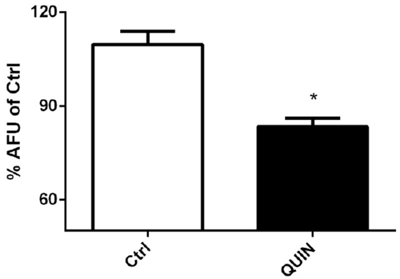
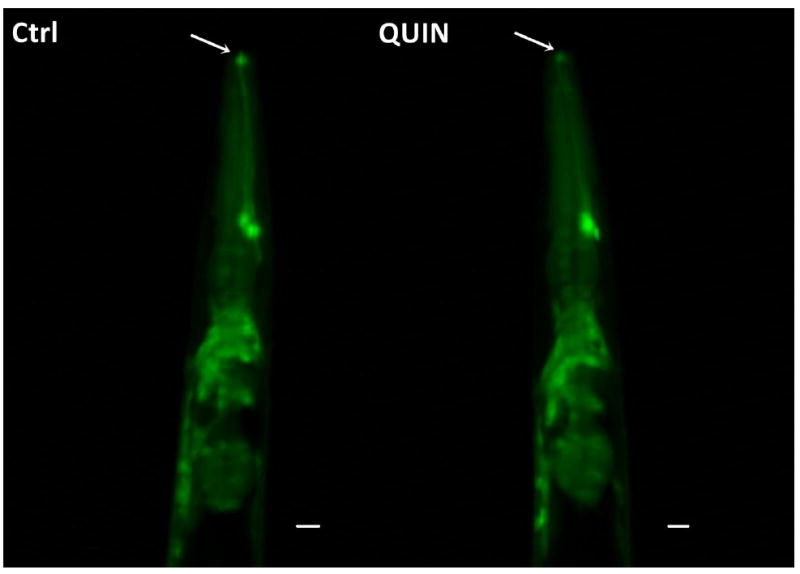
Effect QUIN on neurodegeneration in the OH438 (pan-neuronal∷GFP) strain. The OH438 strain shows all GFP-labeled neurons. A) Quantification of the neurons head fluorescence, indicated neurodegeneration by reducing fluorescence, in the dates expressed as percentage of the arbitrary fluorescence units (AFU) of Ctrl. B) Images the previous region of the worm head. The arrows indicate reduction GFP-fluorescence in neurons. C) GFP-labeled ventral nerve cord without morphological alteration in QUIN-treated worms. Data are derived from three independent assays with 10 worms per group at each experiment (n=30, *p<0.020).
