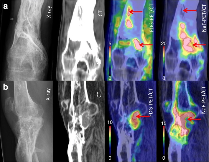
Fig. 1.
Preoperative images (X-ray, CT, FDG-PET/CT, and NaF-PET/CT) of chronic osteomyelitis in distal femur in two different patients. The arrows point at bone segments with high uptake on FDG-PET/CT (“hot spots”) and low uptake on NaF-PET/CT (“cold spots”). The findings are best visualized in coronal views in patient (a), where two sequestra were found, and in sagital views in patient (b), where one sequestrum was found

