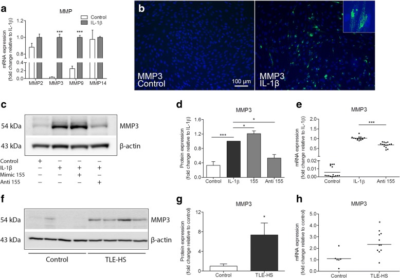
Fig. 1.
Increased MMP3 expression under pathological conditions can be modulated by miR-155 in human astrocytes. a RT-qPCR expression analysis demonstrated increased expression of MMP3 (p < 0.001) and MMP9 (p < 0.001), but not MMP2 and MMP14 mRNA following IL-1β stimulation in human fetal astrocytes. b Immunocytochemistry revealed MMP3 immunoreactive cells 24 h after the stimulation of primary human fetal astrocytes with IL-1β (10 ng/ml), while the immunoreactivity in control cells was not observed; the inset demonstrate a magnified image of cells expressing MMP3 after IL-1β stimulation. c Western blot for MMP3 in human fetal astrocytes. d A semi-quantitative analysis of MMP3 immunoreactivity showed an upregulation of MMP3 after IL-1β stimulation (p < 0.001, n = 5); the cells transfected with miR-155 mimic prior to the stimulation had a further upregulation of MMP3 (p < 0.05, n = 5), and the cells transfected with miR-155 antagomiR showed a decreased expression of MMP3 (p < 0.05, n = 5) compared to non-transfected cells. e RT-qPCR showed decreased MMP3 mRNA expression in the cells transfected with the antagomiR of miR-155 after IL-1β stimulation (p < 0.001, n = 5). f Western blot for MMP3 in TLE-HS specimens compared to autoptic control specimens. g A semi-quantitative analysis of MMP3 immunoreactivity showed an upregulation of MMP3 (p < 0.05) in TLE-HS (n = 6) compared to autoptic control (n = 5). h RT-qPCR analysis showed that MMP3 mRNA was increased (p < 0.05) in a subset of patients with TLE-HS (n = 10) compared to autoptic control (n = 6). Gene expression was represented as normalized to the geometric mean of the expression of two housekeeping genes. Anti = antagomiR; *p < 0.05, **p < 0.01, Mann-Whitney U test, error bars depict the standard error of the mean

