Fig. 4.
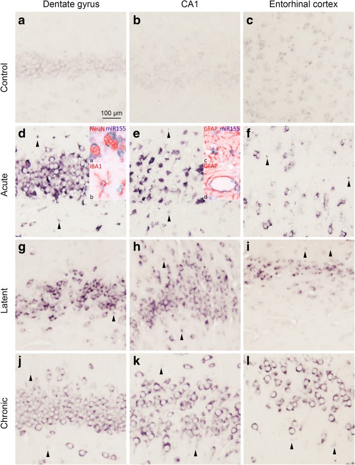
In situ hybridization of miR-155 in the rat TLE model. a–c A weak miR-155 expression was seen in neurons of the control rats in the DG, CA1, and entorhinal cortex (EC). d–l Increased expression compared to control was observed in both neurons and glia of the rats at the acute, latent, and chronic stages. d Co-localization was found with neurons (NeuN; d, inset a), microglia (IBA-1; d, inset b), and astrocytes (GFAP; e, inset c). Additionally, miR-155 expression was seen in blood vessels (e, inset d). Scale bar 100 μm; arrowheads indicate cells with glial morphology

