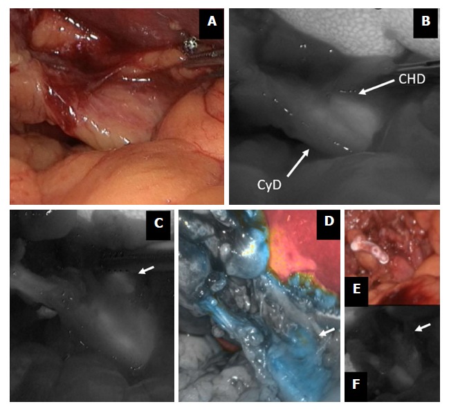
Figure 2.

Indocyanine green-enhanced biliary anatomy. During a difficult cholecystectomy for acute cholecystitis (A), the confluence between the cystic duct (CyD) and the common hepatic duct (CHD) is shown by fluorescence imaging (B); common hepatic duct (arrow) is further visualized before (C and D) and after (E and F) cystic duct division. ICG: Indocyanine green.
