Figure 1.
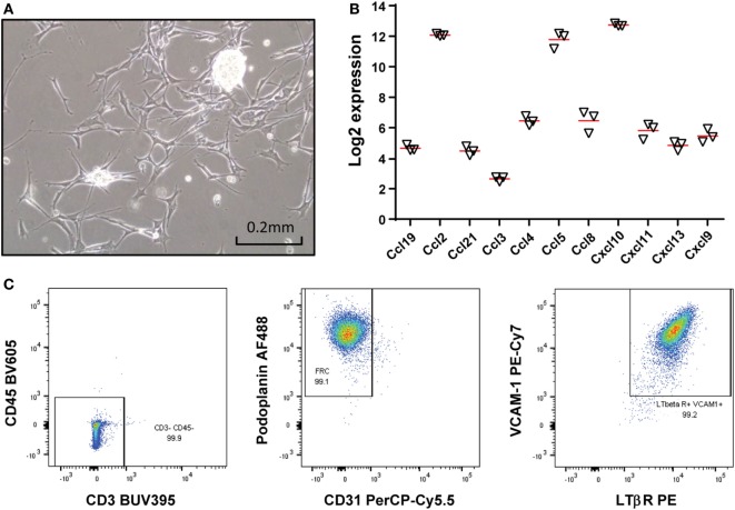
Establishing a lymph node (LN)-derived stromal cell line. (A) A photomicrograph of a LN-derived monoclonal stromal cell line (#2) in culture. Monoclonal cell lines were generated by limiting dilution. Scale bar denotes 0.2 mm. (B) Total RNA was extracted from the stromal cell line (#2) at 3 different passages and mRNA level of indicated 11 chemokines were analyzed by mouse genome arrays. Log2 transformed data were presented and red bars denote the mean. (C) The stromal cell line was stained for CD3, CD45, CD31, podoplanin, LTβ receptor (LTβR), and vascular cell adhesion molecule 1 (VCAM-1), and analyzed by flow cytometry. The majority of the cells are fibroblastic reticular cells with expression of VCAM-1 and LTβR.

